Osteoderm
You can help expand this article with text translated from the corresponding article in German. (June 2014) Click [show] for important translation instructions.
|


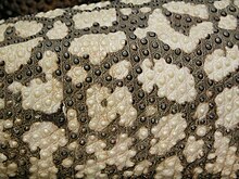
Osteoderms are bony deposits forming scales, plates, or other structures based in the dermis. Osteoderms are found in many groups of extant and extinct reptiles and amphibians, including lizards, crocodilians, frogs, temnospondyls (extinct amphibians), various groups of dinosaurs (most notably ankylosaurs and stegosaurians), phytosaurs, aetosaurs, placodonts, and hupehsuchians (marine reptiles with possible ichthyosaur affinities).
Osteoderms are uncommon in mammals, although they have occurred in many xenarthrans (armadillos and the extinct glyptodonts and mylodontid ground sloths). The heavy, bony osteoderms have evolved independently in many different lineages.[1] The armadillo osteoderm is believed to develop in subcutaneous dermal tissues.[2] These varied structures should be thought of as anatomical analogues, not homologues, and do not necessarily indicate monophyly. The structures are however derived from scutes, common to all classes of amniotes and are an example of what has been termed deep homology.[3] In many cases, osteoderms may function as defensive armor. Osteoderms are composed of bone tissue, and are derived from a scleroblast neural crest cell population during embryonic development of the organism. The scleroblastic neural crest cell population shares some homologous characteristics associated with the dermis.[4] Neural crest cells, through epithelial-to-mesenchymal transition, are thought to contribute to osteoderm development.[2]
The osteoderms of modern crocodilians are heavily vascularized,[5] and can function as both armor and as heat-exchangers,[6] allowing these large reptiles to rapidly raise or lower their temperature. Another function is to neutralize acidosis, caused by being submerged under water for longer periods of time and leading to the accumulation of carbon dioxide in the blood.[7] The calcium and magnesium in the dermal bone will release alkaline ions into the bloodstream, acting as a buffer against acidification of the body fluids.[8]
See also
[edit]References
[edit]- ^ Hill, R.V. (December 2006). "Comparative anatomy and histology of xenarthran osteoderms". Journal of Morphology. 267 (12): 1441–1460. doi:10.1002/jmor.10490. PMID 17103396. S2CID 22294139.
- ^ a b Nasoori, Alireza (2020). "Formation, structure, and function of extra-skeletal bones in mammals". Biological Reviews. 95 (4): 986–1019. doi:10.1111/brv.12597. PMID 32338826. S2CID 216556342.
- ^ Vickaryous, M.K.; Hall, B.K. (April 2008). "Development of the dermal skeleton in Alligator mississippiensis (Archosauria, Crocodylia) with comments on the homology of osteoderms". Journal of Morphology. 269 (4): 398–422. doi:10.1002/jmor.10575. PMID 17960802. S2CID 5927674.
- ^ Vickaryous, Matthew K.; Sire, Jean-Yves (2009-04-01). "The integumentary skeleton of tetrapods: origin, evolution, and development". Journal of Anatomy. 214 (4): 441–464. doi:10.1111/j.1469-7580.2008.01043.x. ISSN 1469-7580. PMC 2736118. PMID 19422424.
- ^ Clarac, F.; Buffrénil, V; Cubo, J; Quilhac, A (2018). "Vascularization in ornamented osteoderms: Physiological implications in ectothermy and amphibious lifestyle in the crocodylomorphs?". Anatomical Record. 301 (1): 175–183. doi:10.1002/ar.23695. PMID 29024422.
- ^ Clarac, F.; Quilhac, A. (2019). "The crocodylian skull and osteoderms: A functional exaptation to ectothermy?" (PDF). Zoology. 132: 31–40. doi:10.1016/j.zool.2018.12.001. PMID 30736927. S2CID 73427451.
- ^ Jackson, DC.; Andrade, D.; Abe, AS. (2003). "Lactate sequestration by osteoderms of the broad-nose caiman, Caiman latirostris, following capture and forced submergence". Journal of Experimental Biology. 206 (Pt 20): 3601–3606. doi:10.1242/jeb.00611. PMID 12966051.
- ^ "Antacid armour key to tetrapod survival". ABC Science. 2012-04-24. Retrieved 6 March 2017.
Further reading
[edit]- Carroll, R. L. 1988. Vertebrate Paleontology and Evolution. W. H. Freeman and Company. ISBN 0-7167-1822-7


 French
 Deutsch