エフリンA5
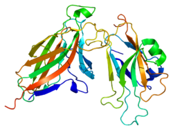
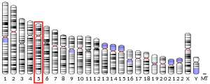
エフリンA5(英: ephrin A5)は、ヒトではEFNA5遺伝子にコードされるタンパク質である[5][6][7]。
エフリンA5はエフリンのAサブクラス(エフリンA)に属するGPIアンカータンパク質であり、EphAサブクラスのEph受容体に結合する。また、エフリンA5はEphB2に結合することも示されている[8]。
成長円錐の生存における逆行性シグナル伝達
[編集]エフリンのユニークな特性の1つは、Eph受容体を発現している細胞へ伝達されるシグナルとは異なるシグナルがエフリンを発現している細胞内へ伝達されるという逆行性シグナル伝達の存在である。エフリンAはGPIアンカーによって細胞膜へ結合しているだけで、エフリンBとは異なり細胞内シグナル伝達ドメインを欠いており、エフリンAによる逆行性シグナル伝達の機構は未解明である。エフリンA5など特定のエフリンAは逆行性シグナル伝達カスケードを開始することが知られており、培養中のマウス脊髄運動ニューロンでは成長円錐の拡大が刺激されることが示されている[9]。エフリンA5による逆行性シグナルはGPI依存的であることが示されており、ホスファチジルイノシトール特異的ホスホリパーゼCによってGPIアンカーを除去することでエフリンA5による成長円錐拡大効果は消失する。EphA受容体は運動ニューロンの成長円錐に対して反対の効果を発揮し、成長円錐の大きさは縮小する。
レチノトピックマップの形成
[編集]エフリンA5は成長円錐の生存を促進し、EphAによるシグナルとは反対の効果を発揮すること、そしてこの効果がエフリンA5による逆行性シグナルによって直接媒介されていることは、軸索誘導に対する重要な示唆をもたらす。すなわち、この現象はEphAを発現している移動中の軸索がエフリンA5を発現している細胞を選択的に回避し、そしておそらくエフリンA5の発現が低い細胞へ向かって移動する機構の説明となる[9]。この機構は、レチノトピックマップの形成時に上丘(SC)内の異なる領域へ網膜神経節細胞(RGC)を誘導する機構と同一である。SCの後側領域で高発現しているエフリンA5は、耳側網膜から移動してきたRGCに発現しているEphAへ結合してその成長円錐の崩壊を誘導することで、RGCの後側領域からの反発とエフリンA5の発現の低い前側領域へ向かう移動を引き起こす[10]。
出典
[編集]- ^ a b c GRCh38: Ensembl release 89: ENSG00000184349 - Ensembl, May 2017
- ^ a b c GRCm38: Ensembl release 89: ENSMUSG00000048915 - Ensembl, May 2017
- ^ Human PubMed Reference:
- ^ Mouse PubMed Reference:
- ^ “The gene encoding LERK-7 (EPLG7, Epl7), a ligand for the Eph-related receptor tyrosine kinases, maps to human chromosome 5 at band q21 and to mouse chromosome 17”. Genomics 35 (2): 376–9. (Sep 1996). doi:10.1006/geno.1996.0371. PMID 8661153.
- ^ “LERK-7: a ligand of the Eph-related kinases is developmentally regulated in the brain”. Cytokine 9 (8): 540–9. (Oct 1997). doi:10.1006/cyto.1997.0199. PMID 9245480.
- ^ “Entrez Gene: EFNA5 ephrin-A5”. 2023年10月1日閲覧。
- ^ “Repelling class discrimination: ephrin-A5 binds to and activates EphB2 receptor signaling”. Nat. Neurosci. 7 (5): 501–9. (May 2004). doi:10.1038/nn1237. PMID 15107857.
- ^ a b “Coexpressed EphA receptors and ephrin-A ligands mediate opposing actions on growth cone navigation from distinct membrane domains”. Cell 121 (1): 127–39. (April 2005). doi:10.1016/j.cell.2005.01.020. PMID 15820684.
- ^ “In vitro guidance of retinal ganglion cell axons by RAGS, a 25 kDa tectal protein related to ligands for Eph receptor tyrosine kinases”. Cell 82 (3): 359–70. (August 1995). doi:10.1016/0092-8674(95)90425-5. PMID 7634326.
関連文献
[編集]- “The ephrins and Eph receptors in neural development”. Annu. Rev. Neurosci. 21: 309–45. (1998). doi:10.1146/annurev.neuro.21.1.309. PMID 9530499.
- Zhou R (1998). “The Eph family receptors and ligands”. Pharmacol. Ther. 77 (3): 151–81. doi:10.1016/S0163-7258(97)00112-5. PMID 9576626.
- “Eph receptors and ephrins: effectors of morphogenesis”. Development 126 (10): 2033–44. (1999). doi:10.1242/dev.126.10.2033. PMID 10207129.
- Wilkinson DG (2000). “Eph receptors and ephrins: regulators of guidance and assembly”. Int. Rev. Cytol.. International Review of Cytology 196: 177–244. doi:10.1016/S0074-7696(00)96005-4. ISBN 978-0-12-364600-2. PMID 10730216.
- “Roles of Eph receptors and ephrins in segmental patterning”. Philos. Trans. R. Soc. Lond. B Biol. Sci. 355 (1399): 993–1002. (2001). doi:10.1098/rstb.2000.0635. PMC 1692797. PMID 11128993.
- Wilkinson DG (2001). “Multiple roles of EPH receptors and ephrins in neural development”. Nat. Rev. Neurosci. 2 (3): 155–64. doi:10.1038/35058515. PMID 11256076.
- Winslow JW; Moran P; Valverde J et al. (1995). “Cloning of AL-1, a ligand for an Eph-related tyrosine kinase receptor involved in axon bundle formation”. Neuron 14 (5): 973–81. doi:10.1016/0896-6273(95)90335-6. PMID 7748564.
- Gale NW; Holland SJ; Valenzuela DM et al. (1996). “Eph receptors and ligands comprise two major specificity subclasses and are reciprocally compartmentalized during embryogenesis”. Neuron 17 (1): 9–19. doi:10.1016/S0896-6273(00)80276-7. PMID 8755474.
- Lackmann M; Mann RJ; Kravets L et al. (1997). “Ligand for EPH-related kinase (LERK) 7 is the preferred high affinity ligand for the HEK receptor”. J. Biol. Chem. 272 (26): 16521–30. doi:10.1074/jbc.272.26.16521. PMID 9195962.
- Ephnomenclaturecommittee (1997). “Unified nomenclature for Eph family receptors and their ligands, the ephrins. Eph Nomenclature Committee”. Cell 90 (3): 403–4. doi:10.1016/S0092-8674(00)80500-0. PMID 9267020.
- Ciossek T; Monschau B; Kremoser C et al. (1998). “Eph receptor-ligand interactions are necessary for guidance of retinal ganglion cell axons in vitro”. Eur. J. Neurosci. 10 (5): 1574–80. doi:10.1046/j.1460-9568.1998.00180.x. PMID 9751130.
- “Ephrin-A binding and EphA receptor expression delineate the matrix compartment of the striatum”. J. Neurosci. 19 (12): 4962–71. (1999). doi:10.1523/JNEUROSCI.19-12-04962.1999. PMC 6782661. PMID 10366629.
- Gerlai R; Shinsky N; Shih A et al. (1999). “Regulation of learning by EphA receptors: a protein targeting study”. J. Neurosci. 19 (21): 9538–49. doi:10.1523/JNEUROSCI.19-21-09538.1999. PMC 6782889. PMID 10531456.
- Davy A; Gale NW; Murray EW et al. (2000). “Compartmentalized signaling by GPI-anchored ephrin-A5 requires the Fyn tyrosine kinase to regulate cellular adhesion”. Genes Dev. 13 (23): 3125–35. doi:10.1101/gad.13.23.3125. PMC 317175. PMID 10601038.


 French
 Deutsch


